In this unit you will learn the basics about the fundamental unit of all living organisms, the cell. We will focus on the following aspects of the cell and what it means in the function of the cell and the rest of the body.
- How did we get here? The cell theory
- What's the difference? Comparison of a prokaryotic cell and a eukaryotic cell
- Why are cells so small? Understanding how surface area and volume affect a cell (math application: surface area, volume, ratios)
- What's inside? Determining the function and affects of the organelles in a cell (Evolution connection: Endosymbiont hypothesis)
- How do I get in? The cell membrane
- How do cells reproduce? Mitosis and Binary Fission
The Cell Theory
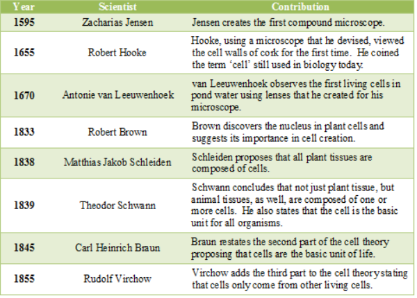
Above is a timeline that describe the cell theory, however it can be boiled down to several basic parts that go even beyond this point. The main three points of the cell theory are
- All living things are compose of cells
- Cells are the basic units of structure and function of all living thing
- New cells are produced from old cells
- All living things are compose of cells
- Cells are the basic units of structure and function of all living thing
- New cells are produced from old cells
- Cells contains hereditary information which is passed from cell to cell during cell division.
- All cells are basically the same in chemical composition.
- All energy flow (metabolism & biochemistry) of life occurs within cells
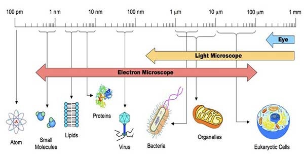
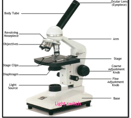
The microscope above is a light microscope. it is typically what most people will use in class and in labs. It is a good microscope because it is relatively inexpensive and it allows you to see living things. However, you cannot see things that are smaller than 100 nanometers (one billionth of a meter). For that, you will need an electron microscope
|
|
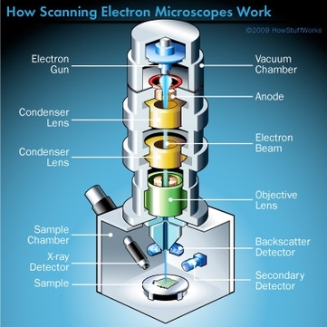
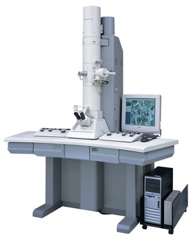
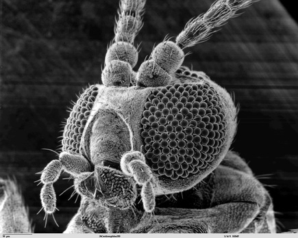
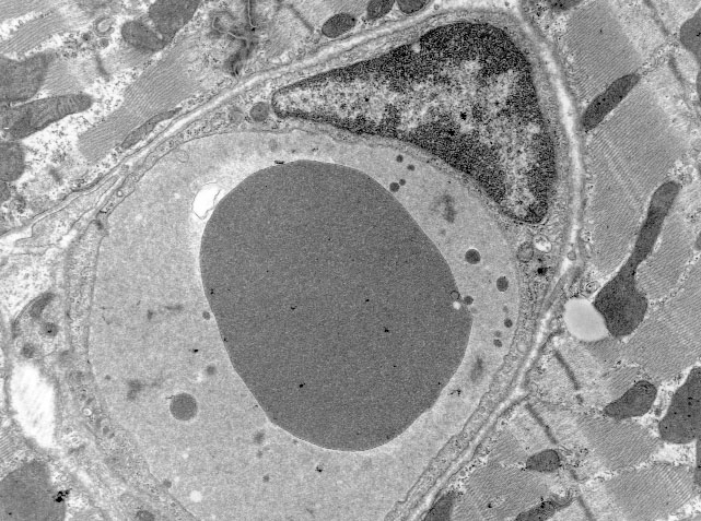
There are two main types of electron microscopes, scanning electron microscopes and transmission electron microscopes. Both are used for viewing small objects, but what they actually are used to view is different. Using a scanning electron microscope, you will shoot electrons at the surface of an object and see only the surface (left, below). However, if you want to see the inside of an object, you will need to but it into thing slices and use a transmission electron microscope (right, below).
|
|
Cells
Cells come in two major groups, Prokaryotes and Eukaryotes. Prokaryotes are often referred to as bacteria, but realize that it is not 100% true as there are more than just bacteria in the Prokaryote group. Most of the time you believe that bacteria are there only to make you sick, but that is also untrue. You have many bacteria in and on you that are necessary for life and actually help you to be healthy. Eukaryotes are typically further broken down into two groups, animal and plant cells. Obviously, you are made of animal cells and plants are made of plant cells. Below, we look at the differences and similarities between the two major groups.
ProkaryotesThere are only a couple of major parts that are found in the prokaryotic cell:
These are the main features, but this does not mean that this is the end of the information about prokaryotes. A major difference between prokaryotes and eukaryotes is the size. Prokaryotes are considerably smaller than eukaryotes, and they do not have any more complex organelles as found in eukaryotes. They have many more structures that help them to be the unique organisms that they are. 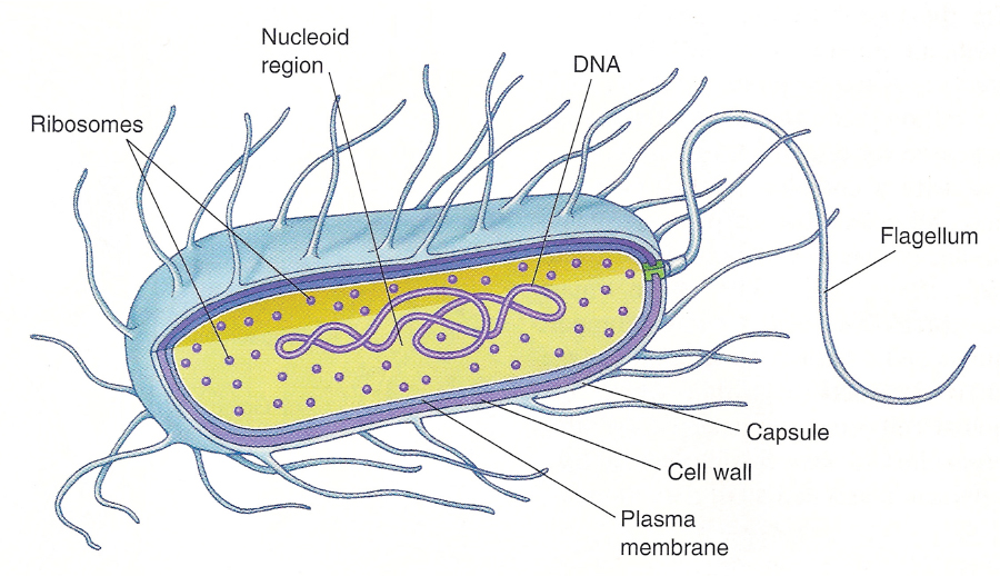
As you can see above, there are the structures that we have already mentioned, but there are some extra structures. You will notice that there is a Flagellum (plural Flagella) that will help with movement of the cell. A cell wall will further protect the cell and fimbriae or pilli that are used to attach to a host cell.
|
EukaryotesEukaryotes have more parts in them than prokaryotes. However, we look at three main differences that eukaryotes have that prokaryotes do not.
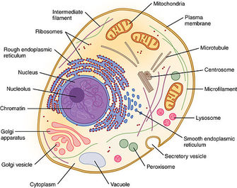
These are the main features, but realize that there are 2 different types of eukaryotic cells, which can contain different organelles themselves- plant cells (below, top) and animal cells (below, bottom). You can also say that because these organisms are a little bit more complex, this also allows them to increase in size because they are able to increase how much energy they get out of their environment. Due to this, they are several times bigger than their prokaryotic cousins. 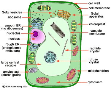
|
Organelles
Before reading everything, here are some video options that you can use that give an overview of the cell, or give specifics about different organelles.
The basics of each organelle that we will be focusing on
- Endoplasmic Reticulum: This is divided into two different groups, each of which has a different function in the cell. Most eukaryotic cells will have both SER and RER.
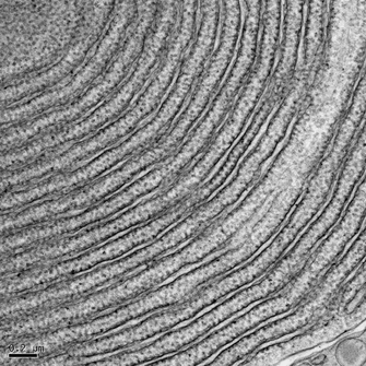
- Smooth Endoplasmic Reticulum- Produces membrane phospholipids, produces sex hormones (ex. testosterone and oestrogen), detoxification of drugs in the liver, storage of calcium to help with muscle contractions, aid liver in releasing glucose in the bloodstream. This type of Endoplasmic Reticulum is characterized by the fact that it does not have any ribosomes attached to it, therefore giving it a smooth appearance when magnified appropriately.
- Rough Endoplasmic Reticulum- Involved in the production of proteins that may become parts of the membrane, messengers or enzymes. This is characterized by the fact that it has ribosomes attached, therefore giving it a rough appearance when viewed under the appropriate magnification.
- Ribosomes- These organelles will synthesize proteins in the cell. They can be found floating freely in the cell or attached to the endoplasmic reticulum.
- Lysosomes- Organelles that are involved in the recycling and breaking down of trash/extra materials in the cell.
- Vacuole- Used for storage in the cell. In plant cells, there is a central vacuole stores water.
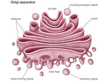
- Golgi apparatus- Assists in the packaging of proteins for the outside of the cell via secretory vesicles
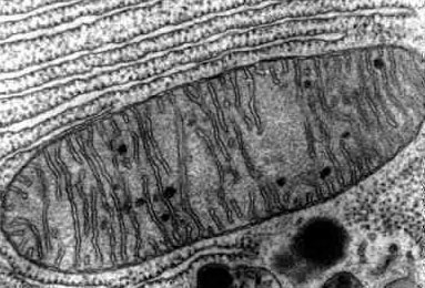
Mitochondria- The primary location for ATP (energy) formation

Nucleus- organelle that acts as the control center of the cell, houses all the DNA
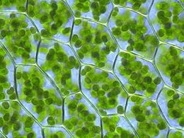
- Chloroplast- Used in photosynthesis
Cell Membrane
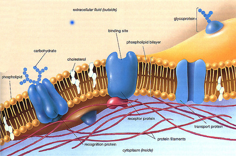
The membrane of the cell is a very important part of the cell. One of its more important functions is to keep objects in the cell as well as keep objects out of the. It also controls what will enter and exit the cell. The model the is used of the cell membrane in called the fluid mosaic model. If you break down the name of the model for the cell membrane it makes sense: fluid for the mobility and mosaic because it is made up of different materials.
Cell Reproduction
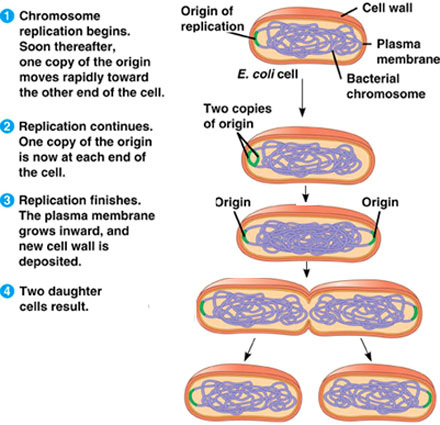
You are surrounded by death. It is everywhere around you. You a surrounded by birth. It is everywhere around you. You really don't think about it, but your body is made up of cells, and these cells are constantly dying (apoptosis- or programmed cell death) and going through reproduction (mitosis). Bacterial cells go through the same thing (binary fission), except that it isn't quite as complicated because prokaryotes are simpler than eukaryotes. So, what is going on in this microscopic circle of life? The simplest answer is that one cell is splitting and creating two copies of itself. However, the process by which a prokaryote with divide is very different from the process by which a eukaryote with divide, hence the discussion of binary fission versus mitosis.
Binary Fission |
Mitosis |
|
Cell Cycle Reproduction in eukaryotic cells requires a longer period of time as it has several more steps than binary fission. More importantly, mitosis is only a small part of the life cycle of a eukaryotic cell. 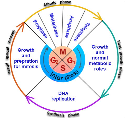
Note that not the entire lifecycle of the cell is mitosis, something a lot of people tend to think is happening. Instead, only a small part of the time is used for mitosis. The rest of the time the cell is in INTERPHASE. During this time, the cell will perform its everyday functions (G1 phase), replicate the DNA (S phase) and grow in preparation for mitosis (G2). During this time, the cell must also go through several checkpoints to make sure that everything is correct. If there are errors in the cell, the process of mitosis should not occur.
M Phase (mitosis) 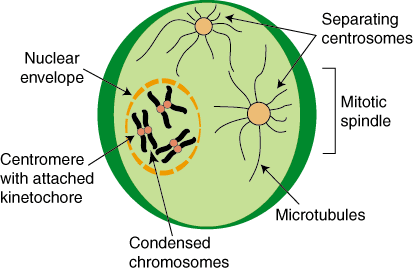
PROPHASE is the first stage of mitosis. In this stage, the chromatin (loose DNA) will condense and become chromosomes. The nucleus will start to fall apart and the centrosomes will start to move towards the poles of the cell.
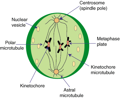
METAPHASE is the next step of mitosis. During this stage the microtubules will attach to the middle of the chromosomes (kinetochore) and arrange the chromosomes so that they are lines up in the middle of the cell. This area is also known as the metaphase plate.
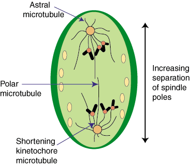
In ANAPHASE, the centrosomes will start to pull the microtubules which will cause the chromosomes to split in half -a lot like a violent version of tug'o'war- , pulling one copy of the DNA to either side of the cell.
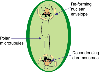
TELOPHASE is the last phase of mitosis. In this stage of mitosis. During this time, the nuclear membrane will start to reform, the cell elongates for splitting (cytokinesis) and the chromosomes start to unpack and go back to the chromatin state.
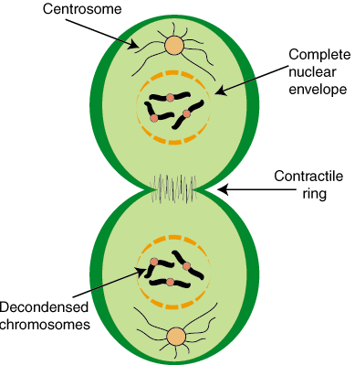
The final step in the circular cell cycle is cytokinesis. At this point, contractile rings made of microtubules will start to constrict and split the cell in two.
|
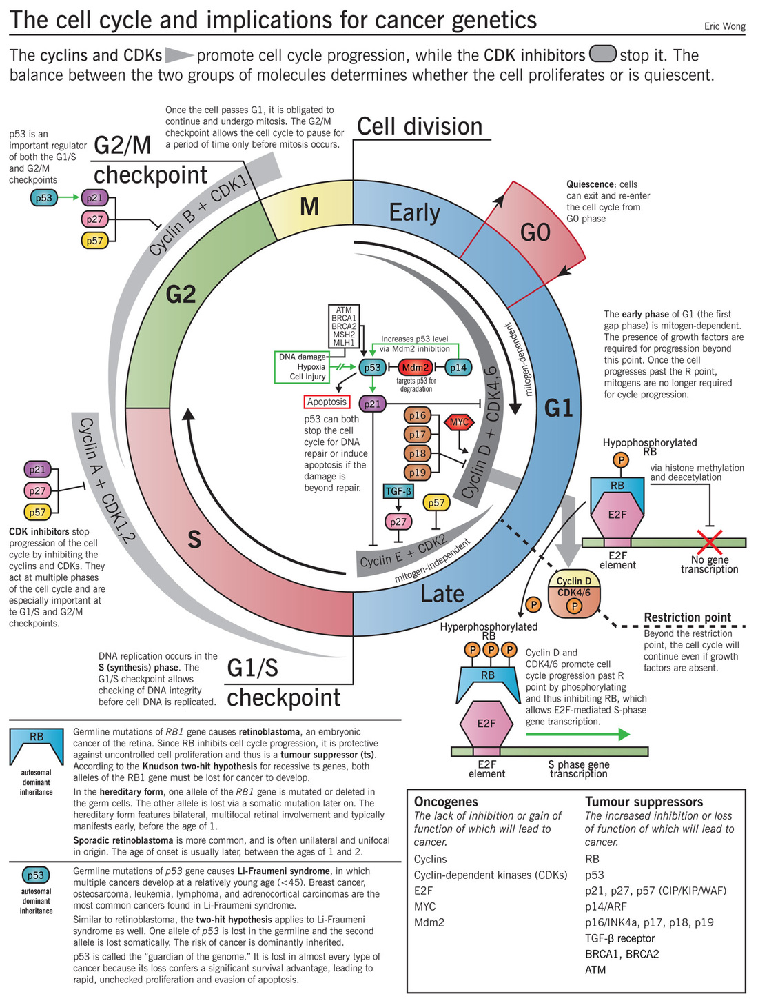
Although there are several checkpoints in the cell cycle. Occasionally something can go wrong. Many times, the body is able to catch the mistake and there destroy the cell or not go through mitosis. However, there are times that the cell will start to uncontrollably reproduce. This is what can lead to cancer and tumors. The diagram below gives some examples of what could happen and where they will occur in the cell cycle.
Images:
http://study.com/cimages/multimages/16/timeline_of_cell_theory..png
http://www.microbiologyinfo.com/wp-content/uploads/2015/05/Size-of-Bacterial-Cells.jpg
http://ayushisinhamicroscopy.weebly.com/uploads/1/4/6/7/14673578/6566165.png
http://uic.igc.gulbenkian.pt/images/microscopy/H-7650.JPG
http://remf.dartmouth.edu/images/insectPart3SEM/image/26cecidomyiidae200.jpg
https://www.med.unc.edu/microscopy/images/electron-microscopy/TEM__BF_1.jpg
https://online.science.psu.edu/sites/default/files/biol011/Fig-3-9-Bacterial-Cell-Organization.jpg
http://waynesword.palomar.edu/images/plant3.gif
http://www.lifesci.sussex.ac.uk/home/Julian_Thorpe/tem3.jpg
http://24.media.tumblr.com/tumblr_lg082dqX4u1qgbt6yo1_500.jpg
http://images.tutorvista.com/cms/images/123/cell-cycle-2.PNG
http://img.sparknotes.com/figures/D/d756b5b73abe2974f3521a828791899f/prophase.gif
http://img.sparknotes.com/figures/D/d756b5b73abe2974f3521a828791899f/metaphase.gif
http://img.sparknotes.com/figures/D/d756b5b73abe2974f3521a828791899f/anaphase.gif
http://img.sparknotes.com/figures/D/d756b5b73abe2974f3521a828791899f/telophase.gif
http://img.sparknotes.com/figures/D/d756b5b73abe2974f3521a828791899f/cytokinesis.gif
http://www.pathophys.org/wp-content/uploads/2012/10/Cell-cycle-copy.jpg
http://quatr.us/biology/cells/pictures/mitochondrion.jpg
http://www.bu.edu/histology/i/20201hoa.jpg
http://alevelnotes.com/content_images/fluid_mosaic.jpg
